Anatomy Of Chest : Male Internal Anatomy Of Chest Photograph By Hank Grebe. Magnetic resonance imaging (mri) utilizes magnet and radio waves to produce diagnostic images. The muscles of the chest develop from the somites found in the mesoderm. It provides protection to vital organs (eg, heart and major vessels, lungs, liver) and provides stability for movement. Learn about each of these muscles, their locations, functional anatomy and exercises for them. As a result of differences in patient age, body habitus, positioning, inspiratory effort, exam technique, and many other factors, normal anatomic structures will vary in appearance on chest radiographs from exam to exam, patient to patient, and even breath to breath.
Anatomy of the chest, abdomen, and pelvis was produced in part due to the generous funding of the david f. Free uk delivery on eligible orders Here, we break down the anatomy of your chest muscles. The pectoralis major and the pectoralis minor, known collectively as your pecs. The epidermis is the outermost layer that provides a protective, waterproof seal over the body.
1 Anatomy Thoracic Key from thoracickey.com Magnetic resonance imaging (mri) utilizes magnet and radio waves to produce diagnostic images. Use the mouse scroll wheel to move the images up and down alternatively use the tiny arrows (>>) on both side of the image to move the images.>>) on both side of the image to move the images. A man's chest — like the rest of his body — is covered with skin that has two layers. Anatomical structures are labeled, providing an invaluable teaching. See chest anatomy stock video clips. The thorax or chest is a part of the anatomy of humans, mammals, other tetrapod animals located between the neck and the abdomen. The first step in understanding thorax anatomy is to find out its boundaries. Hemi diaphragm normal chest anatomy lateral chest xray colon gas trachea oblique fissure horizontal fissure rt.
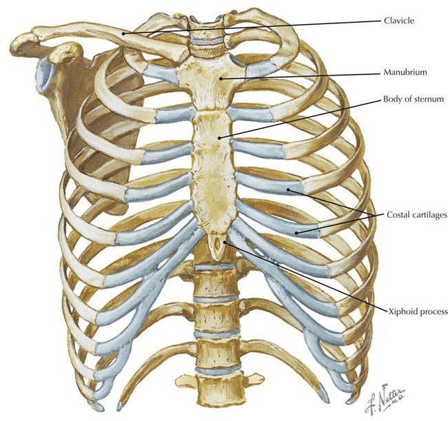
Knowledge of the anatomy of the whole cylinder (ribs, sternum, vertebra, diaphragm, intercostal spaces, and extrathoracic muscles) is therefore not only important in the.
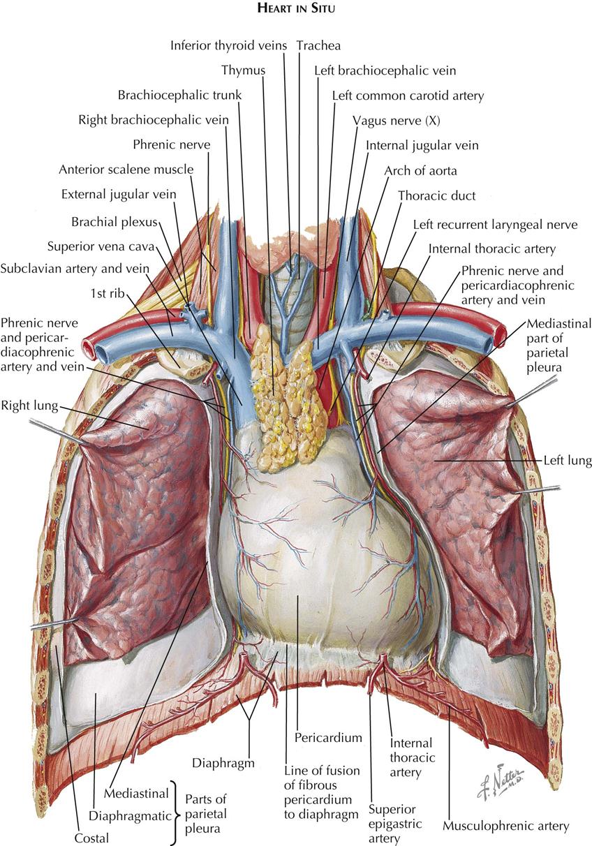
The pectoralis major and the pectoralis minor, known collectively as your pecs. Other important structures, such as the pleura, only become visible when abnormal, and some are not visible at all, such as the phrenic nerve. The chest or thorax is the region between the neck and diaphragm that encloses organs, such as the heart, lungs, esophagus, trachea, and thoracic diaphragm. This page provides an overview of the chest muscle group. Great prices on anatomy of heart. The muscles of the chest develop from the somites found in the mesoderm. Learn about each of these muscles, their locations, functional anatomy and exercises for them. Here, we break down the anatomy of your chest muscles. The pectoralis major, pectoralis minor, serratus anterior and subclavius. Computed tomography (ct) of the chest can detect pathology that may not show up on a conventional chest radiograph(1). Man head and chest anatomy diagram with ghost effect. Anatomy of the chest and the lungs: Principal functions are the protection of internal viscera and an expandable cylinder facilitating variable gas flow into the lungs.
Muscles of the chest and their functions you have two mighty muscles on both sides of your chest: Anatomical structures are labeled, providing an invaluable teaching. The muscles of the chest develop from the somites found in the mesoderm. This atlas is a comprehensive and affordable learning tool for medical students and residents and especially for radiologists and pneumologists. The chest or thorax is the region.
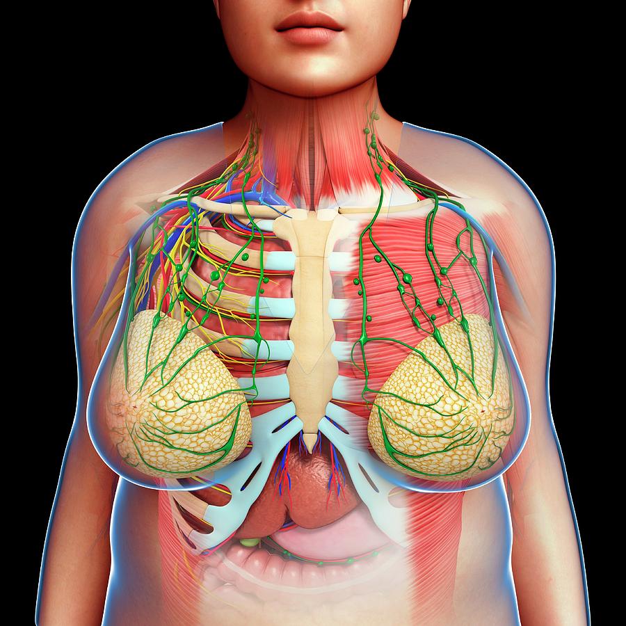
Female Chest Anatomy Photograph By Pixologicstudio Science Photo Library from images.fineartamerica.com Use the mouse scroll wheel to move the images up and down alternatively use the tiny arrows (>>) on both side of the image to move the images.>>) on both side of the image to move the images. Anatomically, the heart is located in the anterior thoracic cavity; Find out more about the individual muscles within the chest anatomy by clicking their respective. Check out anatomy of heart on ebay. This webpage presents the anatomical structures found on hip mri. Find the perfect male chest anatomy stock photo. This thoracic and pulmonary anatomy tool is especially designed for students of anatomy (medical and paramedical studies). Vectors | black & white | cut outs.
Swensen fund for innovation in teaching.
Use the mouse scroll wheel to move the images up and down alternatively use the tiny arrows (>>) on both side of the image to move the images.>>) on both side of the image to move the images. The pectoralis major and the pectoralis minor, known collectively as your pecs. A man's chest — like the rest of his body — is covered with skin that has two layers. This webpage presents the anatomical structures found on hip mri. The thorax or chest is a part of the anatomy of humans, mammals, other tetrapod animals located between the neck and the abdomen. The chest anatomy includes the pectoralis major, pectoralis minor and the serratus anterior. Atlas of ct anatomy of the chest. The pectoral region is located on the anterior chest wall. The circulatory system does most of its work. The first step in understanding thorax anatomy is to find out its boundaries. In insects, crustaceans, and the extinct trilobites, the thorax is one of the three main divisions of the creature's body, each of which is in turn composed of multiple segments. It provides access to ct images in the axial plane, allowing the user to learn and review the lung anatomy interactively. The epidermis is the outermost layer that provides a protective, waterproof seal over the body.
This thoracic and pulmonary anatomy tool is especially designed for students of anatomy (medical and paramedical studies). Atlas of ct anatomy of the chest. Anatomical illustrations of the lungs It provides access to ct images in the axial plane, allowing the user to learn and review the lung anatomy interactively. Computed tomography (ct) of the chest can detect pathology that may not show up on a conventional chest radiograph(1).
Thorax Radiology Key from radiologykey.com It provides access to ct images in the axial plane, allowing the user to learn and review the lung anatomy interactively. Magnetic resonance imaging (mri) utilizes magnet and radio waves to produce diagnostic images. It contains four muscles that exert a force on the upper limb: A man's chest — like the rest of his body — is covered with skin that has two layers. Vectors | black & white | cut outs. No need to register, buy now! The pectoral region is located on the anterior chest wall. The epidermis is the outermost layer that provides a protective, waterproof seal over the body.
How to view the anatomical labels.
Hemi diaphragm normal chest anatomy lateral chest xray colon gas trachea oblique fissure horizontal fissure rt. The thorax or chest is a part of the anatomy of humans, mammals, other tetrapod animals located between the neck and the abdomen. Anatomy of the chest and the lungs: About the 6th week, the somites differentiate into the sclerotomes and the dermatomyotomes. See chest anatomy stock video clips. As a result of differences in patient age, body habitus, positioning, inspiratory effort, exam technique, and many other factors, normal anatomic structures will vary in appearance on chest radiographs from exam to exam, patient to patient, and even breath to breath. Computed tomography (ct) of the chest can detect pathology that may not show up on a conventional chest radiograph(1). Great prices on anatomy of heart. Magnetic resonance imaging (mri) utilizes magnet and radio waves to produce diagnostic images. Anatomical structures are labeled, providing an invaluable teaching. This page provides an overview of the chest muscle group. Man head and chest anatomy diagram with ghost effect. It provides protection to vital organs (eg, heart and major vessels, lungs, liver) and provides stability for movement.
Share this post
0 Response to "Anatomy Of Chest : Male Internal Anatomy Of Chest Photograph By Hank Grebe"


0 Response to "Anatomy Of Chest : Male Internal Anatomy Of Chest Photograph By Hank Grebe"
Post a Comment