Posterior Neck Muscle Diagram / Muscles - Advanced Anatomy 2nd. Ed.. Muscles of the neck posterior triangle prevertebral and lateral muscles. Quickly memorize the terms, phrases and much more. The posterior scalene is the smallest and deepest of the scalene muscles. Click here for a diagram of the the posterior belly of digastric muscle and its relations. It is deeply placed, lying behind sternocleidomastoid.
You've got anterior, middle and posterior scalene muscles. The drawings here present idealized versions of. Muscles of the neck posterior triangle prevertebral and lateral muscles. The scalene muscles are an important part of the anatomy of the neck, with several important structures located between and around them. The masseter muscle originates on the zygomatic arch, and inserts onto.
muscles of head and neck - Anatomy & Physiology 2200 with ... from s3.amazonaws.com I have also done a tutorial on the anterior triangle of the neck, so please watch that if you are interested! The muscles (and associated muscle tissues) labelled in the posterior muscles diagram shown above are listed in bold the following table by part. The posterior scalene is the smallest and deepest of the scalene muscles. Neck and shoulder muscles diagram muscles of neck anterior view dental hygiene pinterest anatomy. This muscle diagram is interactive: Unlike the anterior and middle scalene muscles, it inserts into the second rib. Almost every movement in the body is the outcome of muscle contraction. (supplies deep muscles of the neck).
Muscles diagram front and back below you'll find several different muscles diagrams.
This muscle has three parts. Neck and shoulder muscles diagram muscles of neck anterior view dental hygiene pinterest anatomy. I have also done a tutorial on the anterior triangle of the neck, so please watch that if you are interested! Working in pairs on the left and. You've got anterior, middle and posterior scalene muscles. I'll just flick over to a diagram to show you these, but they originate on the transverse processes of what this muscle does is that it flexes the neck anteriorly. The posterior triangle (or lateral cervical region) is a region of the neck. The healthy posterior neck provides stability and support for the cranium, a flexible and protective spine for movement, balance adaptation. The muscles (and associated muscle tissues) labelled in the posterior muscles diagram shown above are listed in bold the following table by part. Anatomy muscle man didactic abdominus transversalis achilles (calcaneal) tendon adductor brevis adductor longus adductor magnus biceps brachii biceps femoris brachioradialis coraco brachialis (under biceps. Learn vocabulary, terms and more with flashcards, games and other study tools. Posterior muscles in the body. They move the head in every direction, pulling the skull and jaw towards the shoulders, spine, and scapula.
Muscles, connected to bones or internal organs and blood vessels, are in charge for movement. The neck muscles, including the sternocleidomastoid and the trapezius, are responsible for the gross motor movement in the muscular system of the head and neck. Muscle strength is recorded as from 5 to 0 or in a percentage and compared bilaterally whenever possible. Its main function is to aid in biting (starkey, et al., 2011). The healthy posterior neck provides stability and support for the cranium, a flexible and protective spine for movement, balance adaptation.
Posterior Neck Anatomy(1) from www.medicalexhibits.com Your posterior neck muscles are those muscles that lie within the posterior triangle of the neck, beneath that investing layer of fascia, although they are not the only. (supplies deep muscles of the neck). Want to learn more about it? A quiz by hannah purdy. Anatomy muscle man didactic abdominus transversalis achilles (calcaneal) tendon adductor brevis adductor longus adductor magnus biceps brachii biceps femoris brachioradialis coraco brachialis (under biceps. These muscles form a small slip on each side, which is nearly parallel to the posterior belly of the digastric muscle. Quickly memorize the terms, phrases and much more. Almost every movement in the body is the outcome of muscle contraction.
The posterior scalene is the smallest and deepest of the scalene muscles.
There is a printable worksheet available for download here so you can take the quiz with pen and paper. Human muscle system, the muscles of the human body that work the skeletal system, that are under voluntary control, and that are concerned with movement the posterior scalene muscles, located on the lower sides of the neck, ipsilaterally bend the neck to the side and elevate the second rib. It's got a superior, inferior, oblique part and a vertical part. Capitis, of the head even though not mentioned here, this muscle runs up the spine as part of the erector spinae, and so is relevant as part of the posterior cervical muscles. There are anterior muscles diagrams and posterior muscles diagrams. The muscles of the neck anatomical chart shows in beautiful detail the many anterior, posterior, inferior and lateral views of every muscle that makes up the matrix of support for our skull and brain. Advertisements help pay for this website. Thank you for your support. This is an online quiz called muscle diagram: Their main function is contractibility. Posterior border of the sternocleidomastoideus. Fortunately, these muscles, including the posterior neck muscles, can be described in ways that are fairly easy to understand. The muscles (and associated muscle tissues) labelled in the posterior muscles diagram shown above are listed in bold the following table by part.
Involved early gray = muscle: You've got anterior, middle and posterior scalene muscles. All infrahyoid muscles are palpated in a similar way, but may be difficult to distinguish from other infrahyoid muscles; The neck muscles, including the sternocleidomastoid and the trapezius, are responsible for the gross motor movement in the muscular system of the head and neck. The scalenus posterior (posterior scalene) is one of the three scalene muscles in the neck.
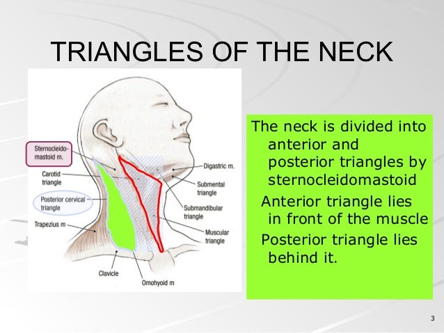
posterior triangle of neck from image.slidesharecdn.com This tutorial covers the muscles of the posterior triangle of the neck as well as the prevertebral and lateral neck muscles. The scalenus posterior (posterior scalene) is one of the three scalene muscles in the neck. Although the sternocleidomastoid muscle begins in the anterior region of the neck, it is considered to be a posterior muscle along with the longissimus capitis muscle, the trapezius muscle, the semispinalis capitis muscle, and the slenius. Union of the sternocleidomastoid and the trapezius muscles at the superior nuchal line of the occipital bone. Advertisements help pay for this website. These muscles form a small slip on each side, which is nearly parallel to the posterior belly of the digastric muscle. Posterior muscles in the body. Working in pairs on the left and.
They move the head in every direction, pulling the skull and jaw towards the shoulders, spine, and scapula.
Muscles, connected to bones or internal organs and blood vessels, are in charge for movement. (supplies deep muscles of the neck). Download scientific diagram | anatomy of short neck muscles. The masseter muscle originates on the zygomatic arch, and inserts onto. This muscle has three parts. Anteriorly by posterior border of sternocleidomastoid muscle. Posterior muscles in the body. Almost every movement in the body is the outcome of muscle contraction. Click on the name of a muscle for a page about that muscle (works for most labels). Capitis, of the head even though not mentioned here, this muscle runs up the spine as part of the erector spinae, and so is relevant as part of the posterior cervical muscles. Lateral flexion, rotation of head to opposite side; Advertisements help pay for this website. This muscle diagram is interactive:
Download scientific diagram | anatomy of short neck muscles neck muscle diagram. Muscles of the neck posterior triangle prevertebral and lateral muscles.


0 Response to "Posterior Neck Muscle Diagram / Muscles - Advanced Anatomy 2nd. Ed."
Post a Comment